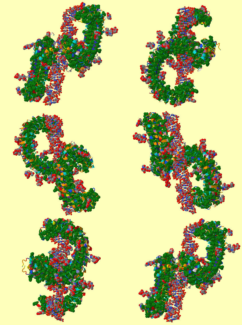

This forms the excited B state, whose emission is blue shifted with respect to A ( Chattoraj et al., 1996). Subsequently, the Thr-203 side chain rotates to donate a hydrogen-bond (HB) to the nascent Tyr-66-anion. The generally accepted model ( Brejc et al., 1997 Palm et al., 1997) for proton migration in photoexcited GFP, suggests that the photodissociated proton of Tyr-66 is transferred, via a water molecule and the OH of Ser-205, to the (presumably anionic) carboxylate group of the buried Glu-222 residue. The Cro assumes a planar (cis) conformation within its rigid cage, and this may be the key to its strong fluorescence. The x-ray data of the wild-type (wt) protein ( Örmo et al., 1996 Yang et al., 1996) show that GFPs possess a unique barrel geometry, consisting of 11 tightly packed β-sheets, with a α-helix carrying the Cro traversing its center. The high RO − quantum yield in GFP contrasts with the denatured protein or the isolated Cro which, like para-phenols ( Schulman et al., 1981), do not fluoresce ( Tsien, 1998 Zimmer, 2002). The GFP Cro appears to follow the same principles ( Scharnagl et al., 1999). It involves intramolecular charge transfer (ICT), from the phenolic oxygen to the aromatic ring system, which is larger for RO − than for the ROH state ( Weller, 1952 Agmon et al., 2002). Indeed, ESPT is a well-known phenomenon in hydroxyaromatics, giving rise to a dramatic decrease in pK a upon electronic excitation ( Weller, 1961 Agmon, 2005). With increasing pH, the amplitude of the latter increases at the expense of the ROH band, showing a clear isosbestic point ( Ward et al., 1982), much like the photoacids that Weller (1952) has investigated 50 years ago. This is evident from the two absorption peaks, at 395 nm for the neutral form (ROH, A state) and 475 nm for the anion (RO −, B state). After photon absorption, it undergoes a reaction of excited-state (ES) proton transfer (ESPT), in which the phenolic hydrogen of Tyr-66 dissociates, leaving behind a brightly fluorescent (green) anion ( Chattoraj et al., 1996 Lossau et al., 1996). Its Cro is synthesized in situ by an autocyclization reaction involving three consecutive amino-acid residues, Ser-65, Tyr-66, and Gly-67. First cloned from the jellyfish Aequorea victoria, it has found extensive application as a biological fluorescence marker. GFP, the “canned candlestick”, is a remarkable solution-phase protein, which is rapidly gaining popularity in molecular biology ( Phillips, 1997 Tsien, 1998 Remington, 2000 Zimmer, 2002).

A plausible conclusion (discussed below) is that GFP is a unique nonmembranal light-driven proton pump, operating according to principles analogous to those of bR. This provides a superb example for the microscopic construction of proton pathways within a protein. Moreover, due to the rigid GFP barrel structure ( Tsien, 1998), all the atomistic “hopping stones” for the translocated proton can be located in the measured x-ray structures ( Örmo et al., 1996 Yang et al., 1996 Brejc et al., 1997 Jain and Ranganathan, 2004), none need to be assumed as in the floppier membranal proton pumps. This work presents surprising findings for the (nonmembranal) green fluorescent protein (GFP), in which well-defined transprotein proton pathways are identified from the x-ray data. To date, proton pathways in nonmembranal proteins have not been thoroughly investigated. In both proteins proton pathways have been identified from the x-ray structure, but due to water disorder, some of the water molecules along these pathways had to be introduced using various computational criteria. This is followed by PT from a buried Asp-96 residue to the Cro, and finally by reprotonation of Asp-96 from the cytoplasmic side. The sequence of proton transfer (PT) events, after photoexcitation of the bR chromophore (Cro), involves conformational change in the Cro and its vicinity, which couples to PT from the Cro to the extracellular surface of the protein. Well-known examples are bacteriorhodopsin (bR) and cytochrome c oxidase (C cO), which convert light or chemical ( ) energy, respectively, into a transmembranal proton gradient, which subsequently drives ATP synthesis ( Wikström, 1998). Proton pumps are a family of membrane proteins that play a pivotal role in the bioenergetics of the cell ( Wikström, 1998 Decoursey, 2003 Ädelroth and Brzezinski, 2004).


 0 kommentar(er)
